Minimally Invasive Craniotomy in Los Angeles
A minimally invasive craniotomy is a specialized neurosurgical procedure designed to treat brain conditions through smaller, more precise openings in the skull. The technique offers the same efficacy as traditional craniotomy but with significantly less disruption to healthy tissue. It is commonly used to access tumors, vascular malformations, and other intracranial abnormalities. At the Global Brain & Spine Institute, brain surgeon Dr. Christopher A. Sarkiss provides minimally invasive craniotomy in Los Angeles, CA.
How Minimally Invasive Craniotomy Works
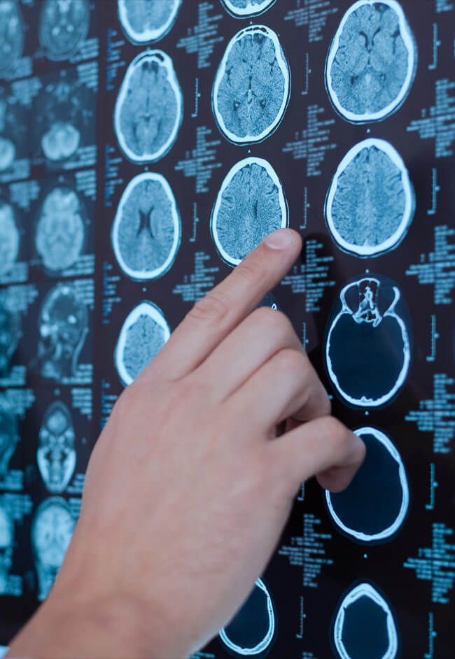
Using high-definition imaging and microsurgical tools, Dr. Sarkiss can navigate delicate brain structures with extreme accuracy. Minimally invasive craniotomy is often performed using endoscopic or keyhole approaches. The precise technique for your surgical procedure will depend on your condition and anatomy. For cases of malignancy, Dr. Sarkiss will remove the tumor with specialized instruments and precision surgical techniques.
Advantages of Minimally Invasive Craniotomy
Key benefits of minimally invasive procedures include shorter hospital stays, reduced postoperative pain, and faster recovery times. Small incisions also mean less visible scarring and a lower risk of complications. Most importantly, a conservative approach can better protect healthy brain tissue.
Schedule a Consultation
If you have been diagnosed with a brain condition requiring surgery, Dr. Sarkiss and his experienced neurosurgical team can assess whether a minimally invasive approach is right for you. Schedule a consultation today to begin working with a top neurosurgeon in Los Angeles, CA.
inimally Invasive Craniotomy FAQs
What conditions can be treated with minimally invasive craniotomy?
Craniotomy procedures are useful across a range of brain conditions and malignancies. Determining the surgical approach will depend not only on the type of condition but also on the size and location of any target lesions or malignancies. Furthermore, condition complexity plays a role in planning, with a minimally invasive approach best suited to well-localized brain conditions.
Dr. Sarkiss may select a minimally invasive craniotomy to treat issues such as:
- Brain tumors
- Meningiomas
- Gliomas
- Brain metastases
- Vascular malformations
- Skull base lesions
Various intracranial abnormalities
Is minimally invasive craniotomy as effective as traditional brain surgery?
How does Dr. Sarkiss determine if I am a candidate?
Procedure planning is a crucial part of any brain surgery or neurosurgery. Doing so entails creating a comprehensive picture of each patient’s health. To this end, Dr. Sarkiss conducts a full assessment and coordinates with other care team members to evaluate:
- Imaging
- Diagnosis
- Symptoms
- Neurological function
- Medical history
- Location of the abnormality
Is minimally invasive craniotomy safe?
Any brain surgery, and any surgery at all, carries some risk. However, minimally invasive techniques are especially designed to minimize these risks, reducing unnecessary trauma and improving outcomes. Surgeon skill and experience also play central roles, including their effective use of advanced imaging and microsurgical tools to enhance accuracy and protect brain structures
How long is the hospital stay after minimally invasive craniotomy?
Most patients can expect a hospital stay of 1 to 4 days, an improvement over the 5 to 7 days of traditional techniques. Though we know you’d like to return home as soon as possible, it is essential to remain in-hospital for inpatient monitoring for a time. This brief period helps ensure the surgery’s success and that there are no concerns before you return home.
What is recovery like after minimally invasive craniotomy?
Following the 1–4 days of hospital recovery, you can spend the next few weeks at home. For about 1 to 3 weeks, patients will need to remain at home and focus on recovery. Fatigue is a common side effect, along with soreness and swelling. Full recovery takes 1–2+ months, with full cognitive and physical form returning after 1 to 3 months post-surgery.
Will I need radiation or chemotherapy after surgery?
Additional treatment may be needed after surgery in select cases. Situations that might require further management include malignant, metastatic, or aggressive tumors. When required, Dr. Sarkiss gladly coordinates care with oncology clinicians to build a combined treatment plan.
How large is the incision for minimally invasive brain surgery?
Incision size can range from 1.4 cm to 3 or 4 cm (about 0.5 to 1.5 inches). Burr hole techniques can further reduce the incision size.
Why is surgeon experience important for minimally invasive craniotomy?
The shortest answer is that experience begets the precision required for a safe, effective procedure. A skilled neurosurgeon must also be capable of making use of the full set of tools available. Microsurgical tools, advanced imaging, even robotic-assisted elements. Lastly, a surgeon must balance smaller access with safe visualization, striking a balance that minimizes damage to surrounding tissues while still fully achieving therapeutic goals.